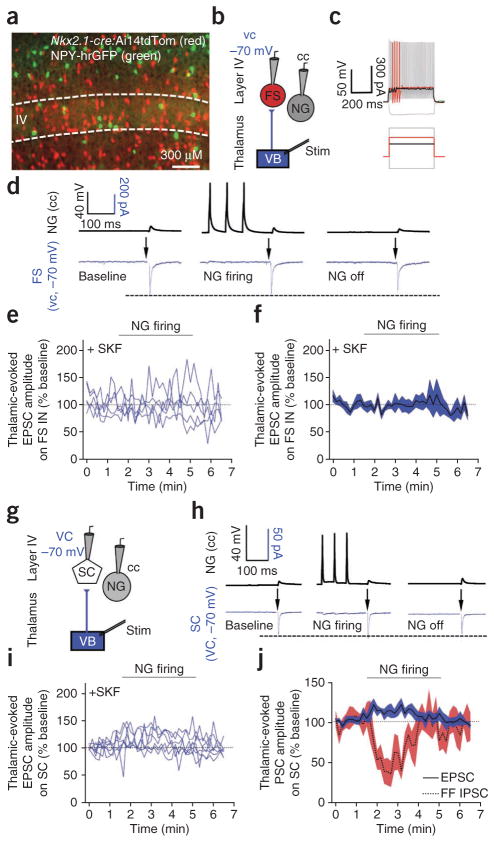
Figure 2.
NGFC activity does not affect thalamic-evoked excitatory input onto layer IV fast-spiking interneurons or stellate cells. (a) Fluorescence image of layer IV in a transgenic mouse expressing NPY-hrGFP and with Nkx2.1-cre activity reported by loxP-flanked tdTomato (Ai14 mouse line). (b) Dual NGFC and fast-spiking interneuron whole-cell recording configuration. (c) Representative firing pattern of fast-spiking interneurons. (d) Single-trace examples of simultaneous current-clamp and voltage-clamp recording in a NGFC (black) and a fast-spiking interneuron (Vh) = −70 mV; blue), respectively. Arrows denote thalamic stimulation. (e,f) Individual (e) and pooled (f) data showing thalamic-evoked EPSC amplitude on fast-spiking interneurons in presence of GAT-1 block by 25 μM SKF-89976A (n = 5). (g) Dual NGFC and stellate cell (SC) whole-cell recording configuration. (h) Single-trace examples of simultaneous current-clamp and voltage-clamp recording in a NGFC (black) and SC (Vh = −70 mV; blue), respectively. Arrows denote thalamic stimulation. (i,j) Individual (i) and pooled (j) data showing EPSC amplitude on SCs at baseline, during and after NGFC firing in presence of GAT-1 block by 25 μM SFK-89976A (n = 7). PSC, postsynaptic current; FF, feed-forward. Red trace in j is a replot of the data shown in Figure 1i. Shaded areas in f and j denote s.e.m. Dotted lines in d and h indicate peak thalamic-evoked EPSC amplitude during baseline

