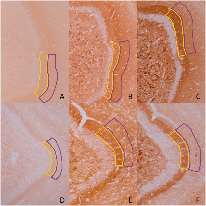
Figure 1.
Representative images of synaptic protein immunoreactivity. Synaptic marker immunohistochemistry from two cases shows distinct staining of the inner molecular layer (highlighted in yellow) and the outer molecular (highlighted in purple) of the hippocampus. (A–C) The relatively healthy outer molecular layer: (A) synaptophysin, (B) SV2 and (C) VGLUT1 with synaptic ratios 1.19, 1.13 and 0.78, respectively (note that VGLUT1 is consistently lower than the other two). In contrast, (D–F) represents an individual with severely reduced outer molecular layer, whose inner molecular layer is preserved: (D) synaptophysin, (E) SV2 and (F) VGLUT1 with synaptic ratios 0.53, 0.74 and 0.56, respectively.

