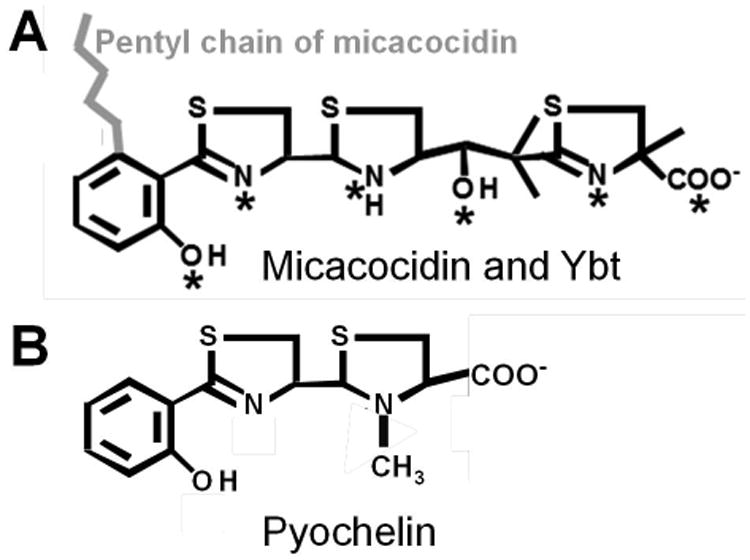
Fig. 1.

The structures of Ybt, micacocidin (A) and pyochelin (B). The asterisks indicate Fe3+ chelation sites in Ybt. The pentyl chain (in grey) of micacocidin is the only structural difference with Ybt (A). Pyochelin (B) lacks the malonyl linker and second thiazoline ring present in YbtA (A).
