
Fig. 6.
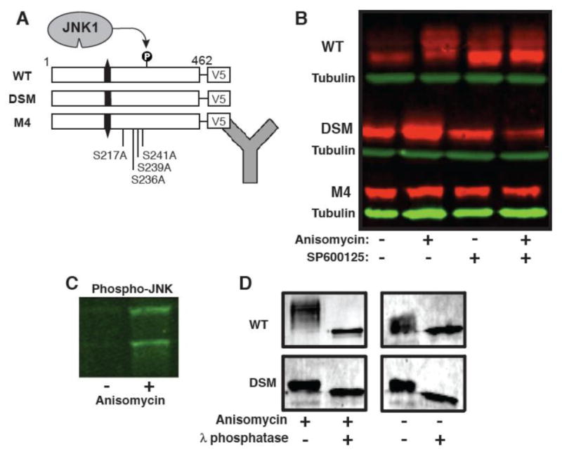
SMTNL2 is an in vivo JNK substrate. A) Wild-type and mutant versions of SMTNL2 were C-terminally-tagged with the V5 epitope and expressed in HEK-293 cells. Then, 24 hours post-transfection, the cells were treated with anisomycin to activate the JNK pathway and/or the JNK inhibitor SP600125. Following this treatment, whole cell protein extracts were prepared and SMTLN2 was visualized by Immunoblotting. B). Anti-V5 immunoblot of whole cell extracts prepared as described in A. C) Anti-phospho-JNK immunoblot of whole cell extracts prepared as described in A. D) WT and DSM-V5 SMTNL2 were immunoprecipitated from 293 cells with or without anisomycin, washed in 1× phosphatase buffer, and then treated lambda protein phosphatase (New England Biolabs) according to the supplied protocol.