Figure 1.
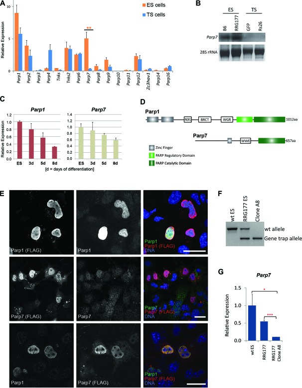
Identification of Parp1 and Parp7 as ES cell-associated genes. (A) RT-qPCR analysis of Parp family members in ES and TS cells. Data of 3–6 independent biological replicates are represented as mean + SEM (**P < 0.01). (B) Northern blot analysis of Parp7 expression in two different ES (B6 and RRG177) and TS (Rs26 and GFP) cell lines. (C) RT-qPCR analysis of Parp1 and Parp7 expression in wildtype (J1 and E14) ES cells over a differentiation time course of 8 days induced by LIF withdrawal and culture on non-adherent plates. (D) Schematic representation of the Parp1 and Parp7 protein domain structure. PADR1 = PADR1 domain; BRCT = BRCA1 C-Terminus domain; WGR = WGR domain; WWE = WWE domain. (E) Immunolocalization of Parp1 and Parp7, detected by confocal microscopy after staining with antibodies against the C-terminal FLAG-tag as well as against the endogenous proteins, showing the nuclear localisation of Parp1 and Parp7. Note that the antibody against endogenous Parp7 is not particularly efficient. Scale bar: 25 μm. (F) Genotyping PCR proving establishment of a homozygously gene-trapped ES cell line at the Parp7 locus. (G) RT-qPCR expression levels of Parp7 in wildtype (wt), Parp7+/− and Parp7-/– ES cells (*P < 0.05, ***P < 0.001).

