
Figure 3.
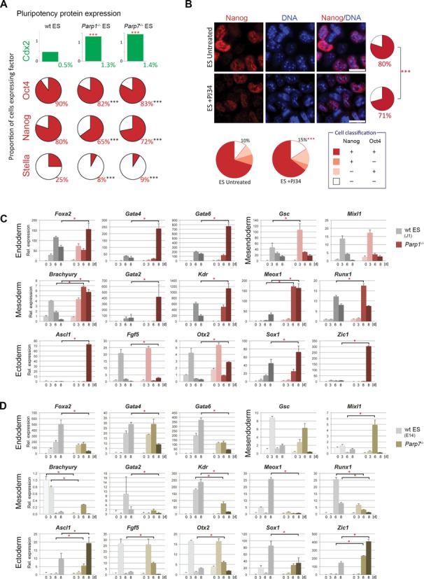
Reduced pluripotency and enhanced differentiation potential of Parp1–/– and Parp7–/– ES cells. (A) Quantification of ES cells stained for Cdx2, Oct4, Nanog and Stella, as shown in Supplementary Figure S1. Cells were classified as positive or negative and compared using a Chi-squared test with the Yates correction. Cell numbers analysed were Cdx2: n = 8544, 5894 and 5415 for wt (J1 and E14), Parp1–/– and Parp7–/–, respectively; Oct4: n = 2034, 2978 and 715; Nanog: n = 1210, 2108 and 1032; Stella: n = 1385, 1555 and 1278; P < 0.001 in all cases. (B) Immunofluorescence staining and quantification for Nanog and Oct4 in wt ES cells cultured for 4 days in ES conditions in either the presence or the absence of the poly-ADP-ribose polymerase inhibitor PJ34. A very similar reduction in pluripotency factor expression is observed. Cell numbers were: Nanog: n = 2476 and 2477; P < 0.001; Oct4: n = 2133 and 1992; P = 0.42; Nanog/Oct4 double staining: n = 1718 and 1045; P < 0.001. Scale bars: 20 μm. (C) and (D) RT-qPCR analysis of marker gene expression levels in embryoid bodies generated by hanging drop culture. Note the skewed differentiation trajectories in both Parp1–/– and Parp7–/– ES cells compared to their background-matched wt control ES cell lines. Parp1-deficient embryoid bodies exhibited a greater variability in size and displayed an overall enhanced speed and/or extent of differentiation towards all embryonic lineages. Parp7 deletion abrogated differentiation towards mesoderm and definitive endoderm (Foxa2, Gata4, Gata6), but overall enhanced ectoderm differentiation.