
Figure 3.
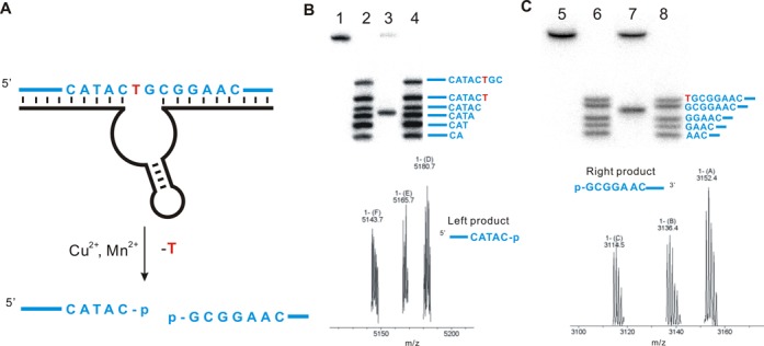
Nucleotide excision of DNA by the DNAzyme F-8. (A) The site of cleavage based on PAGE analysis and mass spectrometry is shown. (B) High-resolution 20% PAGE analysis (upper panel) and mass spectrometry (lower panel) to assign the upstream cleavage fragment generated by F-8. Lane 1, 5′-32P-radiolabeled substrate LS-1; lanes 2 and 4, 5’-32P-radiolabeled oligonucleotide standards based on the substrate LS-1; lane 3, DNA substrate cleaved by F-8 under standard conditions (calcd. 5142.8, found. 5143.7). (C) High-resolution 20% PAGE analysis (upper panel) and mass spectrometry (lower panel) to assign the downstream cleavage fragment generated by F-8. Lane 5, 3’-32P-radiolabeled substrate LS-1+8A (Figure S5.3); lanes 6 and 8, 3’-32P-radiolabeled oligonucleotide standards based on the substrate LS-1+8A; lane 7, DNA substrate cleaved by F-8 under standard conditions (calcd. 3114.5, found 3114.5). The assays indicate that F-8 generates products bearing 3′- and 5′-phosphate termini. Cleavage assays were under standard single-turnover conditions (about 1 μM deoxyribozyme combined with 10 nM substrate).