
Figure 4.
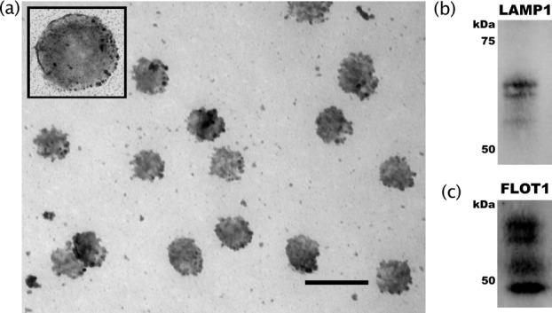
Structural characterization of vesicles as exosomes. Characterization of vesicles released from neurites following potassium-induced depolarization. (a) Glutaraldehyde-fixed samples were sliced and photographed using a JEOL-1200EX transmission electron microscope. Scale bar = 200 nm for main photo, 100 nm for inset. Analysis of particle size using ImageJ software found the average diameter of these vesicles to be 106 ± 7.62 nm. (b) Western blot analysis of exosome markers LAMP1 and (c) Flot1. Each experiment was replicated 3 times and representative blots are depicted.