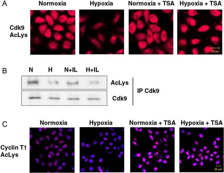
Figure 3.

Hypoxic repression of Cdk9 and Cyclin T1 acetylation. (A) Intracellular distribution of Cdk9 acetylated at lysine residues in response to hypoxia. HeLa cells were pretreated with or without 500 nM TSA for 1 h and subsequently exposed to hypoxia for 1 h. Acetylated Cdk9 was analyzed by P-LISA assay using rabbit anti-AcLys and mouse anti-Cdk9 antibodies. (B) Acetylation of Cdk9 in response to 1 h exposure under normoxic (N) or hypoxic (H) gas mixtures with or without IL-1β. Immunoprecipitated Cdk9 was examined by western blotting with antibodies against acetyl-Lysin (AcLys) or Cdk9. All buffers used in this assay were supplemented with 100 ng/ml TSA to block endogenous HDAC activity. (C) Intracellular distribution of acetylated Cyclin T1 was analyzed by P-LISA assay using rabbit anti-AcLys and goat anti-Cyclin T1 antibodies. HeLa cells were pretreated with or without 500 nM TSA for 1 h followed by exposure to hypoxia for 1 h. Nucleic acids were stained using TOPRO-3 (blue).
