
Figure 2.
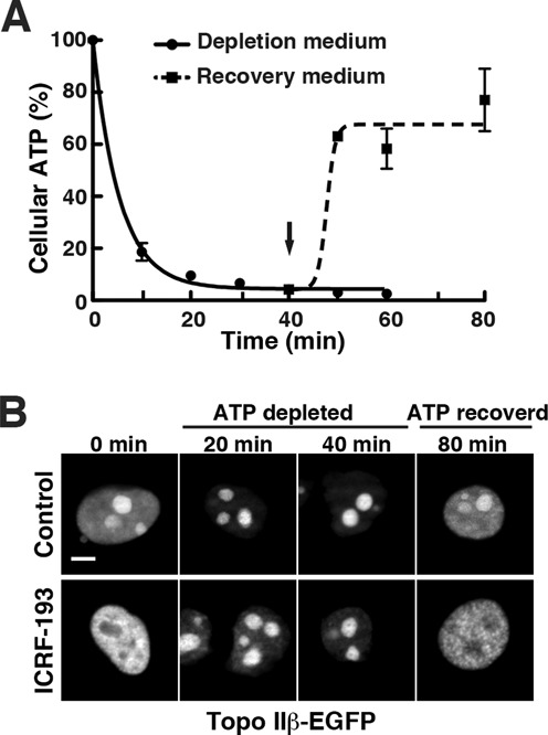
Topo IIβ shuttles between nucleoplasm and nucleoli depending on the cellular content of ATP. At time zero, culture medium was changed to depletion medium containing sodium azide and 2-deoxy-d-glucose. After 40 min, the medium was replaced with normal medium and continued to incubate at 37°C (indicated by arrow). (A) Time course of the relative cellular ATP levels after depletion/recovery treatments. Cells were removed at the time points indicated and cellular ATP was determined by fluorometry and plotted in the graph. Data points represent mean/SD (n = 3). Regression curves were drawn using a software, GraphPad Prism 5 (logistic curve fitting). (B) HEK cells were transfected with pEGFP-top2b and after 24 h, the ATP depletion/recovery procedure was done as in (A). Fluorescence micrographs of nuclei taken at the indicated times are shown in the upper row (control). Shown in the lower row are cells treated with 30 μM ICRF-193 for 15 min at the times indicated. Note that ICRF-induced relocation of topo IIβ does not occur under low ATP condition. Scale bar, 5 μm.