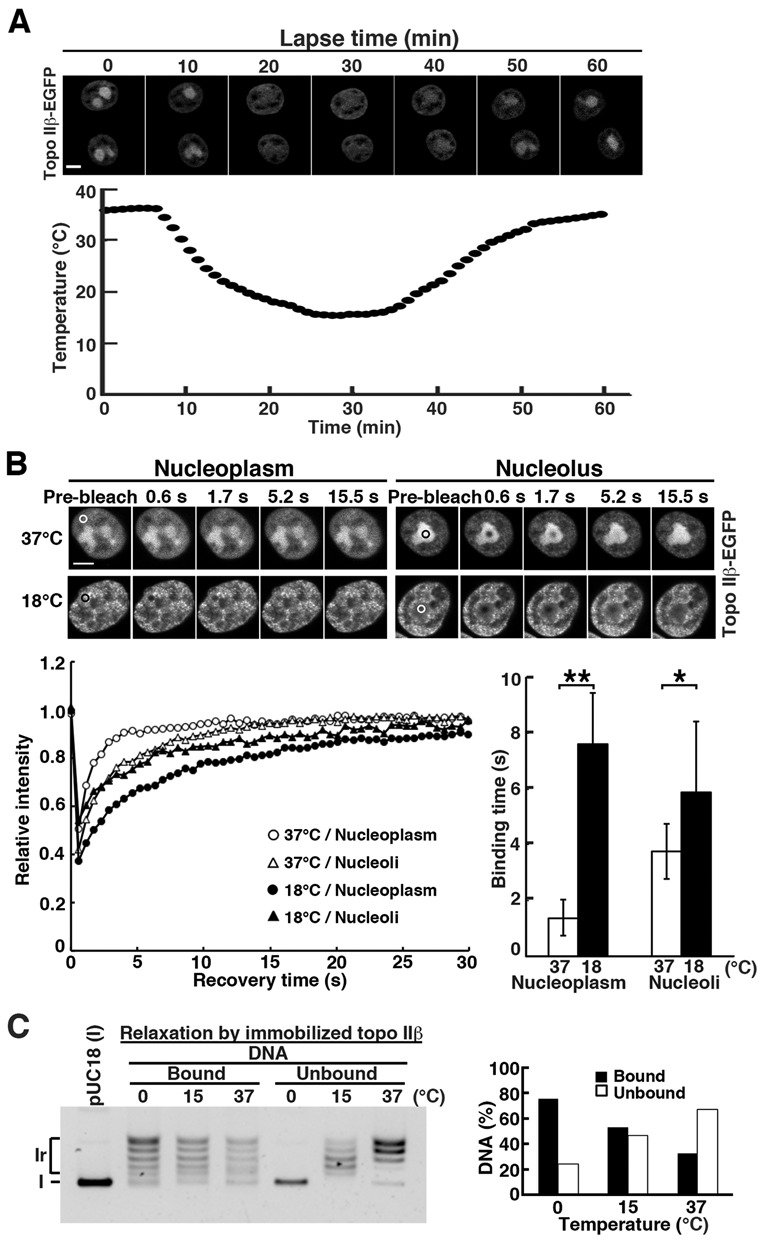
Figure 3.

Cold-induced translocation of topo IIβ from nucleolus to nucleoplasm is accounted for by its lowered catalytic rate. (A) Simultaneous recordings of medium temperature and nuclear distribution of topo IIβ-EGFP expressed in HEK cells. Medium temperature was varied using an on-stage heating/cooling device and monitored by a thermocouple thermometer. Images were recorded at 1-min intervals for 60 min. Scale bar, 5 μm. (B) HEK cells transfected with topo IIβ-EGFP were subjected to FRAP analysis either at 37°C or at 18°C. Fluorescence images were recorded after bleaching the circled areas in nucleoplasm or nucleolus. Representative images and recovery curves (fluorescence relative to pre-bleach) are shown. Plotted in the bar graph are binding times in seconds that were calculated from kinetics data of 24 nuclei for each condition using ImageJ 1.4.6. Bars, mean/SD (n = 24); **P = 1.8 × 10⁻¹⁷, *P = 6 × 10⁻³ by Student's t-test. Scale bar, 5 μm. (C) Temperature dependency of relaxation products in on-bead assay: enzyme-bound versus released DNAs. DNA bands in each lane were quantified by densitometry and relative amounts at each temperature were graphed. Note that the DNA in unbound fraction is fully relaxed at 15°C, but remained supercoiled at 0°C.
