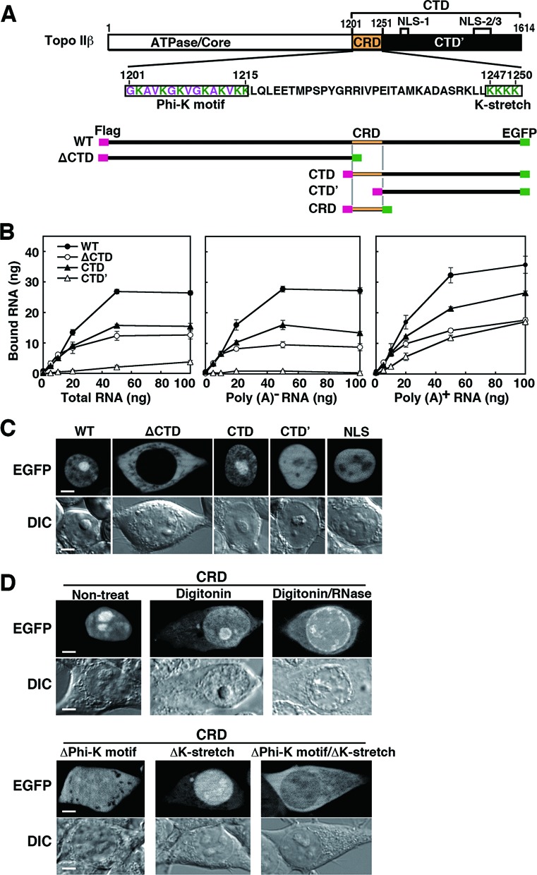
Figure 5.

RNA-binding ability and cellular localization of topo IIβ domain-deletion mutants. (A) Domain diagram of deletion mutants used in this experiment that are dually tagged with Flag/EGFP. Amino acid sequence for CRD (C-terminal regulatory domain) and its subdomains (boxed) are given in the middle. (B) Binding of cellular RNA fractions with topo IIβ deletion mutants immobilized on magnetic beads. Plotted data are expressed in mean with SD (n = 4). (C) Cellular localization of topo IIβ deletion mutants. EGFP images are shown along with differential interference contrast (DIC) images. Images for the SV40 NLS (PKKKRKV) cloned in pEGFP-N1 are put on the right as a control. Scale bars, 5 μm. (D) Localization of CRD in intact, digitonin-treated and digitonin/RNase-treated cells (upper panel). The subdomains boxed in A were further deleted from CRD and their cellular localizations were examined (lower panel). EGFP images are shown along with DIC images. Scale bars, 5 μm.
