
FIGURE 3.
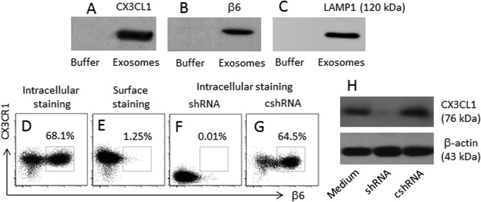
B cells capture HUVEC-derived αvβ6. Exosomes were purified from the HUVEC culture supernatant. Proteins were extracted from the exosomes. A–C, the immune blots indicate the proteins of CX3CL1 (A), αvβ6 (B), and LAMP1 (C) in the extracts of exosomes. D–G, BC-3 cells were cultured in the basal chambers of Transwells. HUVEC were cultured in the inserts for 24 h. The BC-3 cells were analyzed by flow cytometry. The dot plots indicate the frequency of αvβ6+ CX3CR1+ B cells; the treatment is denoted above each subpanel. H, the CX3CL1 gene in HUVEC of F are knocked down by RNAi. shRNA, HUVEC were treated with shRNA of CX3CL1. cshRNA, control shRNA. The data represent three separate experiments.