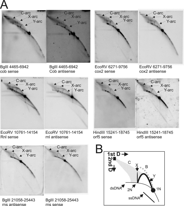
FIGURE 2.
Topological analyses of the C. parapsilosis mtDNA by two-dimensional NAGE shows significant formation of recombination intermediates. A, two-dimensional radiographs as examples from both coding regions to show Y-, X-, and C-arcs present in all of the coding regions. B, interpretation of two-dimensional AGE radiographs where: C, cloud-arc; X, X-arc; Y, Y-arc; 1N, non-replicated restriction fragment; 2N, almost fully replicated intermediates. The dotted gray line named B indicates the expected pattern that would be observed by bubble-shaped intermediates. For positions of restriction enzyme cleavage and probe positions refer to Fig. 1A.

