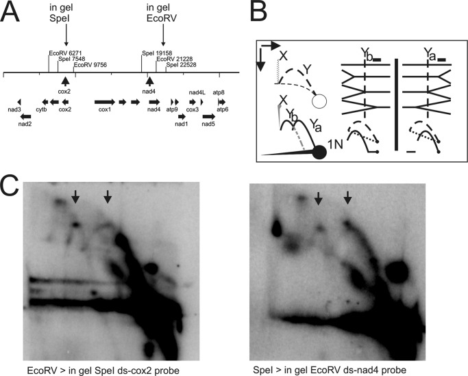
FIGURE 3.
Fork direction two-dimensional AGE of C. parapsilosis mtDNA. A, restriction map indicating cleavage sites for the initial mtDNA digest and showing the position of the site used for in gel cleavage of replication intermediates. B, interpretation of potentially forming Y-arcs depending on the polarity of the analyzed replication fork. Dashed lines indicate the original two-dimensional NAGE pattern, heavy lines depict the detectable patterns after in gelo treatment. The position of the probe is indicated by a black filled box above the Y-structures. The vertical dotted line crossing the Y-structures mimics the position of in gelo cleavage. C, mtDNA digested with EcoRV or SpeI was separated on a first-dimension gel, digested with SpeI or EcoRV in gelo and then separated on a second-dimension gel. Y-arc patterns of both possible fork polarities are seen on both radiographs, thus showing that replication forks pass the fragments bi-directionally.

