FIGURE 9.
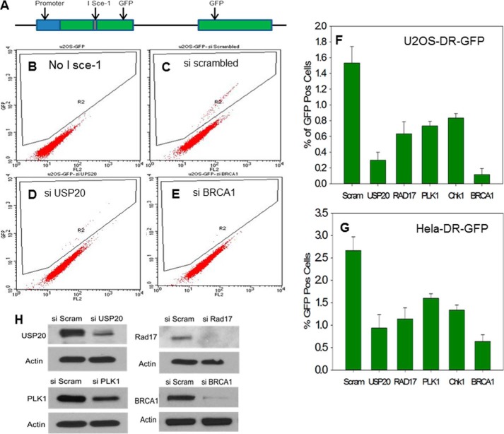
Defective HR in cells depleted of USP20. A, depiction of the HR substrate. Details of the assay are provided in the text. B, U2OS-DR-GFP cells in the absence of the restriction enzyme I Sce-1. No GFP-positive cells are detected. C, GFP-positive cells are observed after treatment with scrambled siRNA and I Sce-1. D, lower percentage of GFP cells when USP20 is depleted. E, dramatic decrease of GFP-positive cells after the depletion of the key HR protein BRCA1. F, representation of percentage of GFP-positive cells after the depletion of USP20. Rad17, PLK1, Chk1, and BRCA1 in U2OS, and (G) Hela cells. The means of three experiments with the standard deviations are shown. H, extent of the protein depletion is shown.

