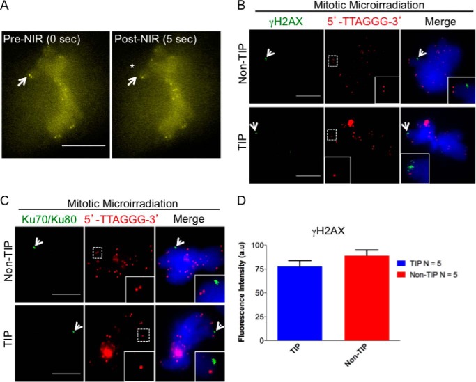
FIGURE 1.
Laser-induced damage activates the DDR in mitotic chromosomes. A, mitotic PtK2 cell stably expressing YFP-TRF2. Images were taken before microirradiation (pre-NIR) and at 5 s postmicroirradiation (post-NIR). White arrows point to a single telomere before (0 s) and after laser microirradiation (5 s) (also indicated with an asterisk). B, arrows point to the accumulation of γ-H2AX (green) at damaged non-TIP and TIP sites at 5 min after laser microirradiation. Telomeres are detected by the 5′-TTAGGG-3′ FISH probe (red). C, the arrow points to the accumulation of Ku70-Ku80 complex at non-TIP and TIP damage sites. The insets show 2-fold magnification of the images in the dotted boxes. Scale bar, 10 μm. D, quantitative measurements (mean ± S.D. (error bars), p = 0.08) after 120-s accumulation of γ-H2AX at TIP and non-TIP damage sites. Statistics are for five independent values (n = 5). a.u., arbitrary units.

