
FIGURE 5.
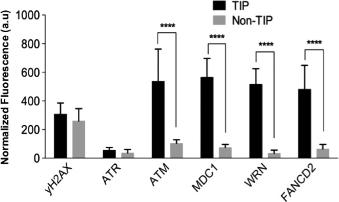
ATM, MDC1, FANCD2, and WRN are preferentially recruited to damage TIPs. Cells were damaged at TIPs and non-TIPs and were fixed at 15 min after damage induction. Cells were subjected to immunofluorescent staining using antibodies specific for γ-H2AX and the indicated DDR factors as in Figs. 3 and 4. Fluorescent signals were quantified by ImageJ software, normalized by the fluorescence signal of the background. n = 10 cells. Two-tailed, unpaired Student's t test was used to obtain p values. a.u., arbitrary units. Error bars, S.D.