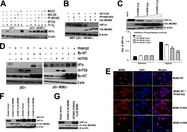
FIGURE 6.
MDM2 regulates PI3K induced hypoxic degradation of HIF1α. A, serum-starved p53−/− or p53−/− MDM2−/− MEFs (4 h) were treated with the proteosome inhibitor 10 μm MG132 for 5 min before being pulsed for 30 min with, the pan-PI3K inhibitors, 25 μm SF1126, and 500 nm PF4691502 and BEZ, followed by IGF stimulation and hypoxia for 4 h. Whole cell extracts were prepared for HIF1α blots. The results support a requirement for MDM2 for PI3K inhibitors effects on hypoxic HIF1α. In the absence of MDM2, inhibitor has no effect on HIF1α under hypoxic conditions. B, MEF p53−/− MDM2−/− cell lines were transfected with 10 μg of HA-MDM2 plasmid using Lipofectamine Plus transfection reagent. 48 h after transfection, cells were serum-starved, followed by treatment with, the pan-PI3K inhibitors, 25 μm SF1126, and 500 nm PF4691502 followed by IGF stimulation and hypoxia for 4 h and whole cell lysate preparation and Western blot analysis. C, LN229-HRE-AP cell lines transfected with PRK5-HA-MDM2 plasmid (6, 8, and 10 μg) or a control plasmid (10 μg) were serum-starved for 4 h and exposed to hypoxia for 4 h, followed by preparation of whole cell extracts (upper panel). LN229-HRE-AP cell lines transfected with PRK5-HA MDM2 plasmid (6, 8, and 10 μg) and control plasmid (10 μg) were exposed to normoxia or hypoxia for 24 h followed by lysate preparation and alkaline phosphatase activity as described before (lower panel). D, p53−/− (left panel) and p53−/− MDM2−/− (right panel) MEFs were transfected with 10 μg of HA-PTEN or Myr-AKT plasmids using Lipofectamine Plus transfection reagent. 48 h after transfection, cells were serum-starved for 4 h before being pulsed for 30 min with 500 nm of PF 4691502 followed by IGF stimulation and hypoxia for 4 h. Whole cell extracts were prepared for HIF1α, HA, and AKT blots. E, localization of MDM2 characterized by confocal microscopy. p53−/− MDM2−/− MEFs were transfected with 1 μg of FLAG-S166A/S166E-MDM2 or HA-WT MDM2 plasmid. Cells were serum-starved for 4 h after 48 h of transfection followed by treatment with 500 nm of PF4691502 for 30 min and were then processed for confocal microscopy as described under “Experimental Procedures.” F, p53−/− MDM2−/− cells were transfected with 10 μg of S166A or S166E MDM2 plasmids. 48 h after transfection, cells were serum-starved, followed by IGF treatment and pulse with 10 μm MG132 followed by hypoxia for 4 h and whole cell extract preparation. These lysates were run on SDS-PAGE, transferred on nitrocellulose membrane, and probed for HIF1α and FLAG tag. G, p53−/− MDM2−/− cells were transfected with 10 μg of S166A or C464A,S166A double mutant MDM2 plasmids. 48 h after transfection cells were serum-starved, followed by IGF treatment and hypoxia for 4 h and whole cell extract preparation. These lysates were run on SDS-PAGE, and proteins were transferred to a nitrocellulose membrane and probed for HIF1α and FLAG tag antibody.

