
FIGURE 4.
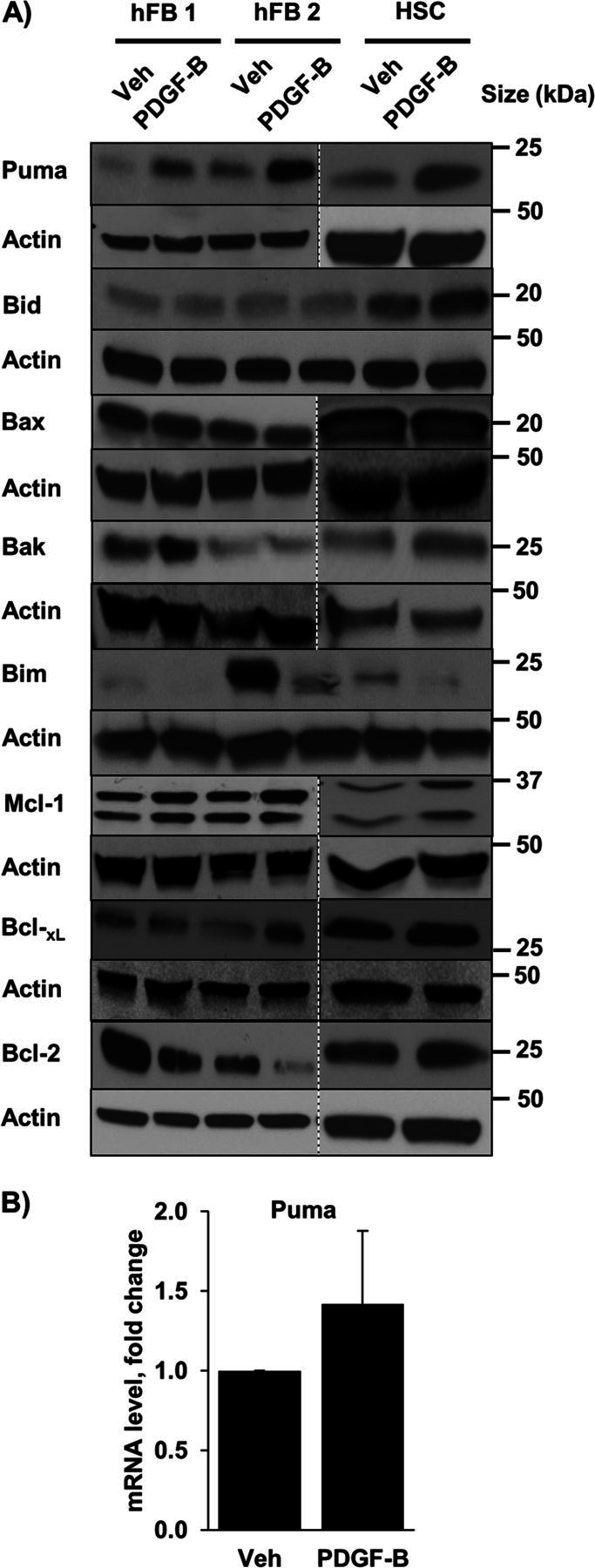
PDGF increases cellular Puma levels. A, whole cell lysates were prepared from quiescent hFB and HSC treated with vehicle (Veh) or PDGF-B for 24 h. Cell lysates were subject to immunoblot analysis of the Bcl-2 family of proteins. Actin was used as a loading control. Except where indicated by dashed lines, all lanes were adjacent on the membranes. B, expression of Puma was assessed by quantitative real-time-PCR in HSC. Means ± S.E. are depicted for n = 3.