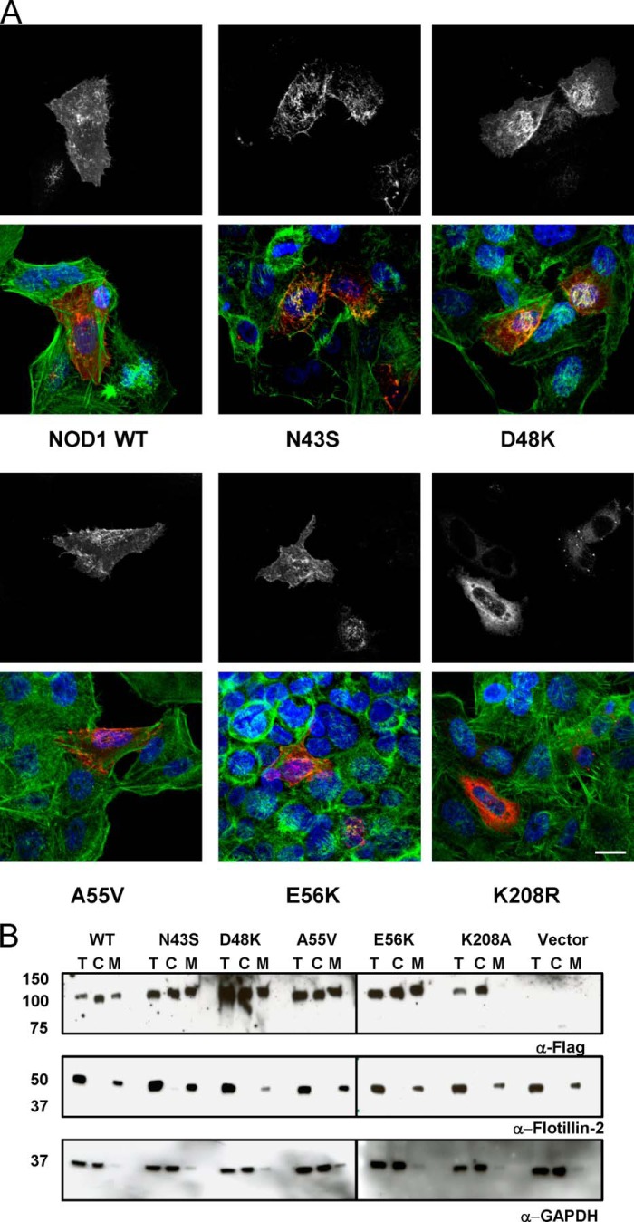
FIGURE 4.
The cellular localization of NOD1 SNPs is comparable with that of wild-type NOD1. A, immunofluorescence studies of FLAG-tagged NOD1 constructs transiently transfected into HeLa cells using FuGENE 6. NOD1 proteins (white in top panels and red in bottom panels) were detected with mouse anti-FLAG M2 and goat anti-mouse Alexa Fluor 546 antibodies. DNA (blue) was stained with DAPI, and actin (green) was stained with phalloidin-FITC. B, subcellular fractionation of FLAG-NOD1 constructs was performed using a Subcellular Fractionation kit. Samples were probed with antibodies against FLAG to detect NOD1, Flotillin-2 to detect the membrane fraction, and GAPDH to detect the cytoplasmic fraction. T, total lysate; C, cytoplasmic fraction; M, membrane fraction. Images are representative of at least two (A) or three (B) independent experiments. Scale bar, 10 μm.

