FIGURE 6.
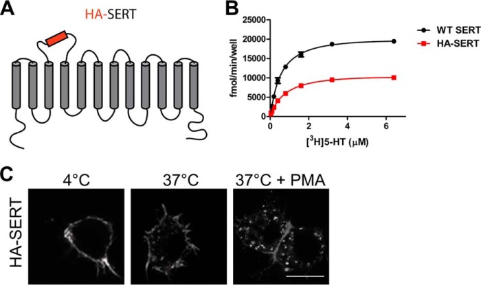
HA-tagged SERT expressed at the cell membrane is functional and is internalized constitutively and after PMA treatment. A, topology diagram of HA-SERT with the HA epitope inserted in the EL2. B, kinetic measurements of specific 5-[3H]HT uptake in CAD cells expressing WT SERT (Km, 0.587 μm; Vmax, 21,771 fmol/min/well) or HA-SERT (Km, 0.727 μm; Vmax, 11,430 fmol/min/well). The curve is representative of three independent experiments. C, antibody feeding internalization assay. Confocal images of CAD cells expressing HA-SERT detected by Alexa568-conjugated HA.11. After incubation with the antibody at 18 °C, cells were either kept at 4 °C (no trafficking) or at 37 °C for 30 min to allow internalization. When indicated, 1 μm PMA was added during the 37 °C incubation. Scale bar,10 μm.

