FIGURE 1.
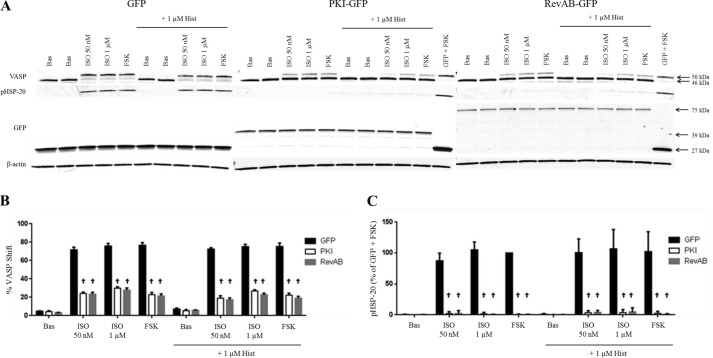
β-Agonist regulation of VASP and phospho-HSP-20 and the effect of PKA inhibition. GFP-, PKI-GFP-, or RevAB-expressing HASM cells were plated into 12-well plates and stimulated with vehicle (Bas), ISO (50 nm or 1 μm) or FSK (100 μm) ± HIST (1 μm) as described under “Experimental Procedures.” After 5 min of stimulation, cell lysates were harvested, and expression levels of various proteins were assessed by immunoblotting. Representative blots (A) are shown. Band intensities for VASP phosphorylation and phospho-HSP-20 were quantified as described under “Experimental Procedures,” adjusted for quantified β-actin levels and presented in graph form in B and C, respectively. VASP bands present as either a 46- or 50-kDa species, the latter representing VASP in which Ser-157 is phosphorylated by PKA. Expression of GFP in all three lines was confirmed by Western blot using anti-GFP antibody. HSP-20 phosphorylation has also been shown previously to be PKA-dependent. Quantified phospho-HSP20 was normalized to the GFP + FSK value. Data are mean ± S.E. †, p < 0.0001 PKI/RevAB versus GFP.

