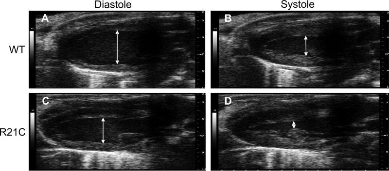
FIGURE 1.
Representative live ECHO images of old (12 months) WT and R21C mice at diastole and systole. A and C, long axis parasternal view of left internal ventricular diameters during diastole in WT (A) and R21C mice (C). B and D, left internal ventricular diameters during systole in WT (B) and R21C mice (D).

