
FIGURE 3.
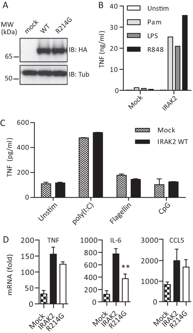
Irak2-deficient macrophages reconstituted with human IRAK2 R214G show altered cytokine responses upon stimulation with TLR2 ligands. A, anti-HA immunoblot analysis of whole cell lysates of mock, HA-IRAK2 WT, and HA-R214G reconstituted macrophages (see “Experimental Procedures”). B and C, reconstitution of Irak2-deficient murine macrophages with human IRAK2 is functional as assessed by TNF secretion analyzed by ELISA (triplicates ± S.D. (error bars)) for a 16-h stimulation with 1 μg/ml Pam2CSK4 (light gray), 0.05 μg/ml LPS (dark gray), and 1 μg/ml R848 (black) (B) but not with 10 μg/ml poly(I:C) (TLR3 ligand), 50 ng/ml flagellin (TLR5 ligand), and 0.5 μm CpG 1668 (TLR9 ligand) (C) or 50 ng/ml recombinant murine IL-1β (not shown). One of four (B) or two (C) representative experiments is shown. D, TNF, IL-6, and CCL5 mRNA induction in mock (checkered), WT human HA-IRAK2 (black), and R214G (white) reconstituted macrophages upon stimulation with Pam2CSK4 for 6 h, relative to unstimulated (triplicates ± S.D. of one representative experiment of two independent experiments shown).