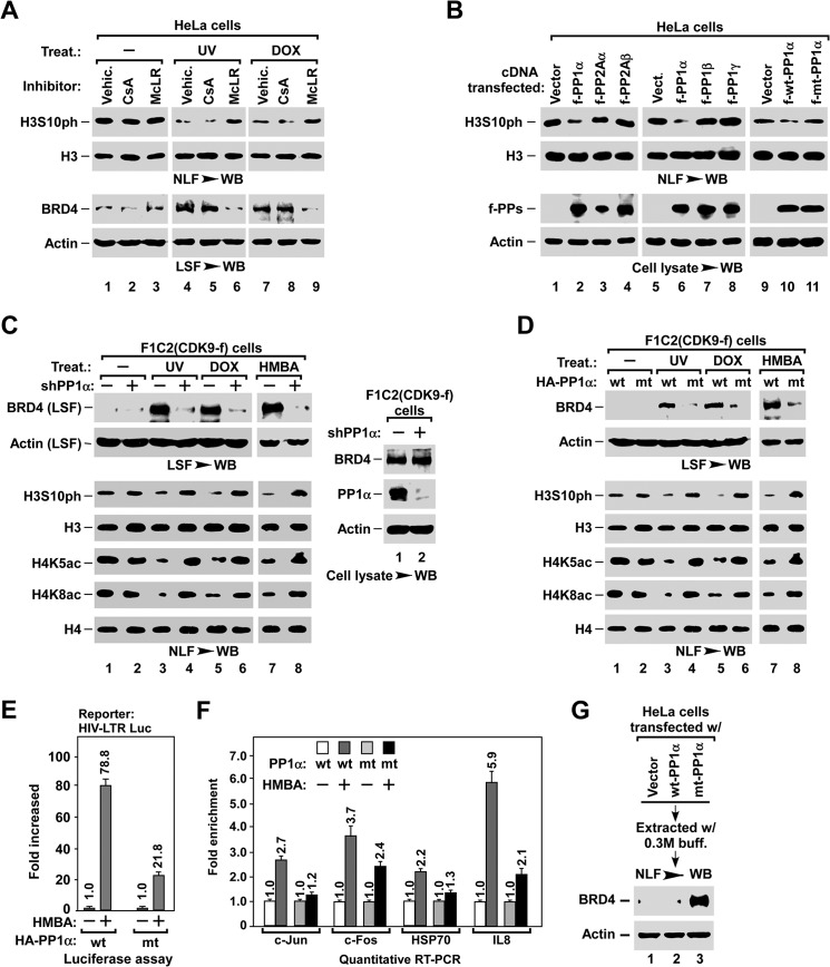
FIGURE 4.
PP1α is required for stress-induced BRD4 release and inducible gene expression. A, effect of phosphatase inhibitors on stress-induced H3S10ph dephosphorylation (top) and BRD4 release (bottom). LSF and NLF were prepared from HeLa cells with the indicated pre-treatment and treatment, and analyzed by Western blot (WB). McLR, PP1/PP2A inhibitor microcystin LR; CsA, PP2B inhibitor cyclosporin A. B, effect of exogenous expression of PP1 and PP2A isoforms on H3S10ph dephosphorylation in NLF (top). The levels of expressed protein phosphatases in cell lysates (bottom) were determined by Western blot (WB). C and D, inhibitory effect of knocking down PP1α (C) or overexpressing H66N mt-PP1α (D) on stress-induced BRD4 release and histone modifications. LSF and NLF were prepared from F1C2(CDK9-f) cells with the indicated lentiviral infection and treatment, and analyzed by Westen blot. E and F, inhibitory effect of overexpressing H66N mt-PP1α on HMBA-induced expression of inducible genes. HIV-LTR-Luc cells with the indicated lentiviral infection and HMBA treatment were subjected to luciferase assay (E) and qRT-PCR analysis (F) as described in the legend to Fig. 3, E and F. G, effect of overexpressing H66N mt-PP1α on the association of BRD4 with chromatin. NLF was prepared from D0.3 M high salt buffer-extracted nuclei of HeLa cells with the indicated lentiviral infection, and analyzed for the remaining BRD4 by Western blot.

