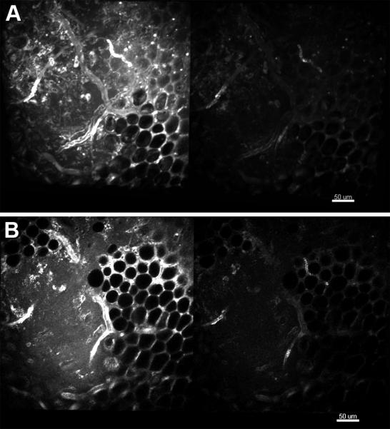
Figure 2.

Comparison of the compact TED collection efficiency and that of the objective alone for imaging fat and mixed tissue in murine skeletal muscle near the knee. A. Maximum intensity projection for the first 25 microns collected with the parabola and objective (left) and the objective alone (right). B. Single imaging plane from approximately 20 microns into the image stack depicted in A. Details of the imaging parameters can be found in “Materials and Methods”.
