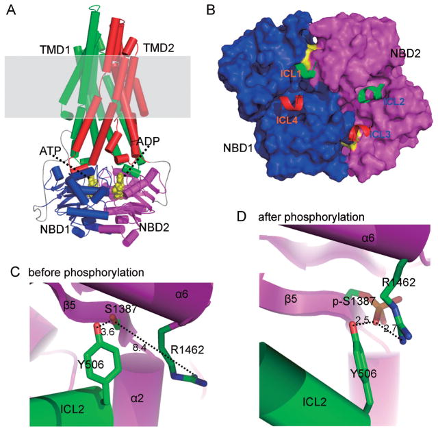
Fig. 2.
Model of SUR2B core domains. A: SUR2B core domains (TMD1, 2 and NBD1, 2) are modeled using SAV1866 as a template. Shaded region is plasmic membrane. B: The interface between TMDs and NBDs are highlighted. Intracellular loop-1 (ICL1) of TMD1 physically interacts with NBD1 and NBD2. ICL2 of TMD1 only interacts with NBD2. Thus TMD1 mainly interact with NBD2. Similarly, TMD2 mainly interacts with NBD1. C and D: The three critical residues involved in PKA phosphorylation are highlighted. Note the side chain of Arg1462 is far from phosphorlation residue Ser1387 before phosphorylation. It is attracted by phosphorylated Ser1387 (p-Ser1387) after PKA phosphorylation and forms a tight triad with p-Ser1387 and Tyr506. The figure is modified fromJournal of Biological Chemistry with permission [16].

