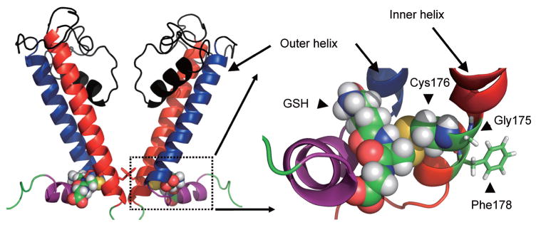
Fig. 3.

Structural modeling of Kir6.1 protein with the incorporation of GSH. The overall structural model of two opposing Kir6.1 monomers (out of four for clarity) was displayed. Boxed area was enlarged and showed the GSH associated area. The GSH moiety occupies a space between the inner and outer helix. The addition of GSH therefore impairs the movement of inner helix, which is necessary for the channel opening. The figure is modified fromJournal of Biological Chemistry with permission [77].
