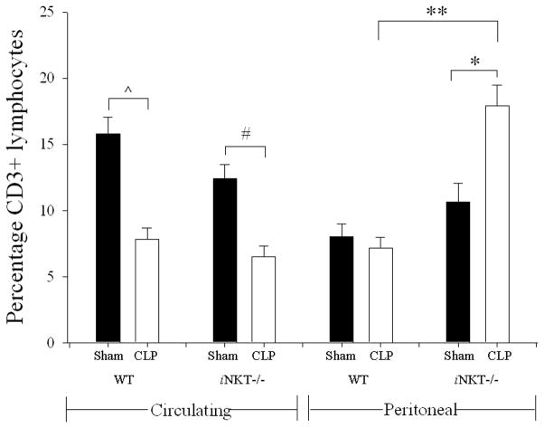
Figure 1. Percentage of peripheral circulating and peritoneal CD3+-lymphocytes in wild type and iNKT−/− mice.
Following CLP, there was a decline in percentage of circulating CD3+-lymphocytes in both wild type (^ p<0.001) and iNKT−/− (# p=0.001) mice. With respect to the peritoneal cavity following CLP compared to Sham, there was no difference in wild type CD3+-lymphocytes; however iNKT−/− mice displayed an increase in percentage of peritoneal CD3+-lymphocytes (* p<0.001). Notably there was no difference in sham peritoneal CD3+-lymphocytes comparing wild type and iNKT−/− mice. However with respect to CLP, peritoneal CD3+ Lymphocytes were significantly greater in iNKT−/− mice compared to wild type (** p<0.001).

