
Figure 2. iNKT-cell profile within a) liver, b) blood, and c) peritoneum following CLP comparing wild type versus PD1−/− mice.
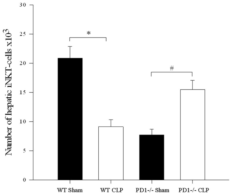
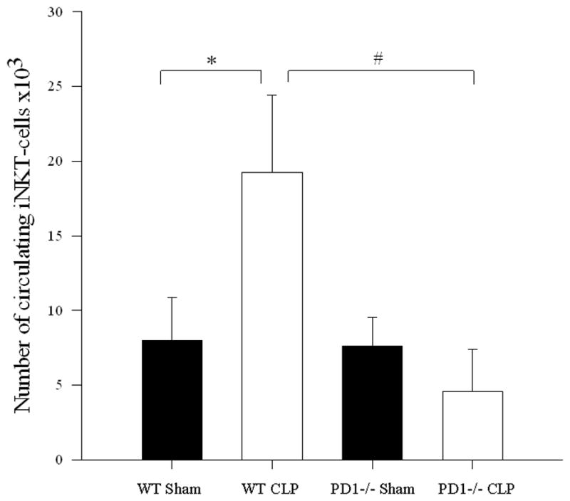
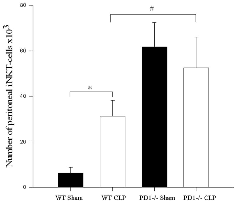
a) Following CLP, there was a decrease in hepatic iNKT-cells in wild type mice (* p<0.001), but an increase in hepatic iNKT-cells in PD1−/− mice (# p=0.004). b) In the peripheral circulation, although wild type mice displayed an increase in circulating iNKT-cells (* p<0.001), there was no difference in the number of circulating iNKT-cells between sham and CLP within PD1−/− mice. PD-1−/− mice had significantly fewer circulating iNKT-cells than wild type mice following CLP (# p<0.001). c) Within the peritoneal cavity, wild type mice displayed an increase in iNKT-cells following CLP (* p<0.001), whereas there was no change in the number of iNKT-cells in PD1−/− mice. Notably PD-1−/− mice had greater peritoneal iNKT-cells compared to wild type following CLP (# p<0.001).