
Figure 3. Effect of bortezomib on ER stress and unfolded protein response (UPR) in infected and uninfected cancer cells.
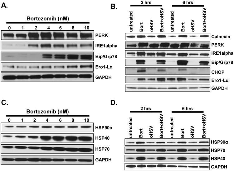
A) Dose dependent induction of ER stress in U251T3 cells. Shown are immunoblots of cell lysates from U251T3 glioma cells treated with bortezomib for 16 hours probed for expression of the indicated proteins. B) ER stress is not increased in cells treated with both bortezomib and oHSV compared to bortezomib only treated cells. Shown are immune blots of the U251T3 cells treated with/without bortezomib (12nM) for 16 hours prior to 34.5ENVE infection at an MOI of 0.01 or 1. Cells were harvested 2 and 6 hours post infection, and cell lysates were probed with antibodies against ER stress related pathway (Calnexin, PERK, IRE1alpha, Bip/GRP78, CHOP, and Ero1-Lα) GAPDH was used as a loading control. C) Dose dependent induction of HSP40, 70 and 90α in U251T3 cancer cells. Shown are immunoblots of cell lysates from the indicated cells treated with bortezomib for 16 hours probed for expression of the indicated proteins. D) Bortezomib pretreatment induced HSPs expression in uninfected and oHSV-infected U251T3 cells. U251T3 cells treated with/without bortezomib were infected with 34.5ENVE (MOI = 1) and cells were harvested 2 and 6 hours post infection. Cell lysates were probed with antibodies against HSP40, HSP70, and HSP90α. GAPDH was used as a loading control.