
Figure 6. Histological characterization of tumor tissues.
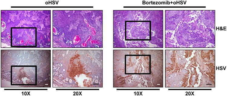
Athymic nude mice were subcutaneously implanted with CAL27 and treated as in Fig. 5. Mice were injected intra-tumorally twice with 1×10⁵ pfu of oHSV every other day. Three days later from second virus injection, tumors were harvested and sectioned. Data shown are representative images of H&E- and HSV-stained tumor sections. Original magnification = × 10 and × 20, as indicated. Scale bar = 100 μm.