
Figure 3. Electrically stimulated Ca2+ transients measured with GCaMP2.2Low and GCaMP2.2Low-FKBP12.6.
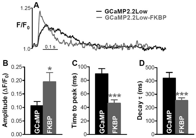
(A) Representative recordings in myocytes expressing GCaMP2.2Low and GCaMP2.2Low-FKBP12.6. (B-D) Amplitude (B), time-to-peak (C) and decay time (D) of Ca2+ transients reported by GCaMP2.2Low (GCaMP) and GCaMP2.2Low-FKBP12.6 (FKBP). Data are the mean±SEM of 10 (GCaMP2.2Low) and 13 (GCaMP2.2Low-FKBP12.6) separate experiments.