
Fig. 3.
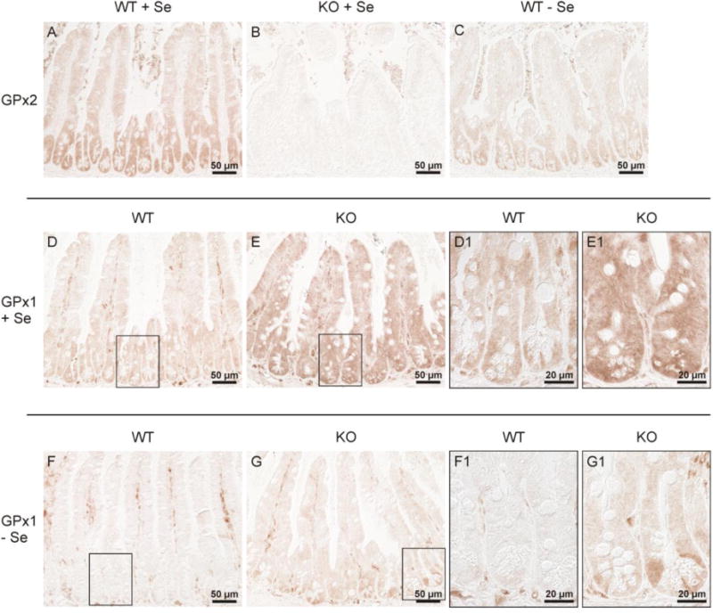
GPx1 and GPx2 in the ileum of wild-type and GPx2 KO mice.
Immunohistochemical GPx2 staining in the ileum of Se-adequate wild-type (A) and GPx2 KO (B) mice shows a similar distribution of GPx2 in the ileum as in the colon: increasing levels from villi towards crypts (A), confirmation of the absence of GPx2 in GPx2 KO mice (B), and a still visible expression under selenium-restriction (C). Staining for GPx1 reveals a drastic increase in GPx2 KO (E, E1) compared to wild-type (D, D1) selenium-adequate mice. GPx1 is almost completely decreased in selenium-poor wild-type (F, F1), but remains slightly up-regulated in selenium-poor GPx2 KO (G, G1) mice, especially in the crypts. The higher magnification of the area marked in the respective overview is labelled by 1. Shown are representative slices of each group stained simultaneously.