
Fig. 5.
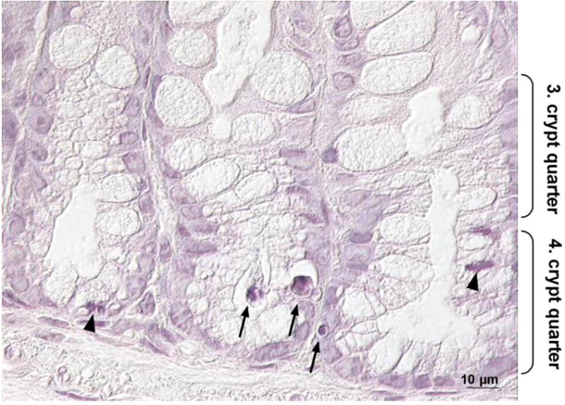
Apoptotic and mitotic cells in the colon of Se-adequate GPx2 KO mice. Slices of colon tissues from 20 weeks old animals were stained with hematoxylin to identify apoptotic and mitotic cells. A representative photograph is shown with apoptotic cells marked by arrows and mitotic cells by arrowheads.