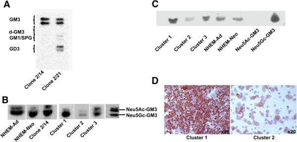
Figure 4.

Correlation between Neu5Gc-GM3 and melanoma cell malignancy. (A) HPTLC separation of gangliosides of two cell clones (2/14 and 2/21) isolated from a single patient. (B) HPTLC separation of Neu5Ac-GM3 and Neu5Gc-GM3 in NHEM-Ad, NHEM-Neo, clone 2/14, and in melanoma cell lines representative of cluster 1 (L6), cluster 2 (L34), cluster 3 (L4). (C) 14 F7 antibody immunostaining of gangliosides separated by HPTLC (NHEM-Ad, NHEM-Neo, L6 representative of cluster 1, L34 representative of cluster 2, L4 representative of cluster 3. (D) Immunohistochemical analysis of cytospin preparations of L6 representative of cluster 1 and L34 representative of cluster 2, stained with 14 F7 antibody. Original magnification, x20 (Axiovert 100). Similar results were obtained for other melanoma cell lines according to the clustering.
