
Fig. 7.
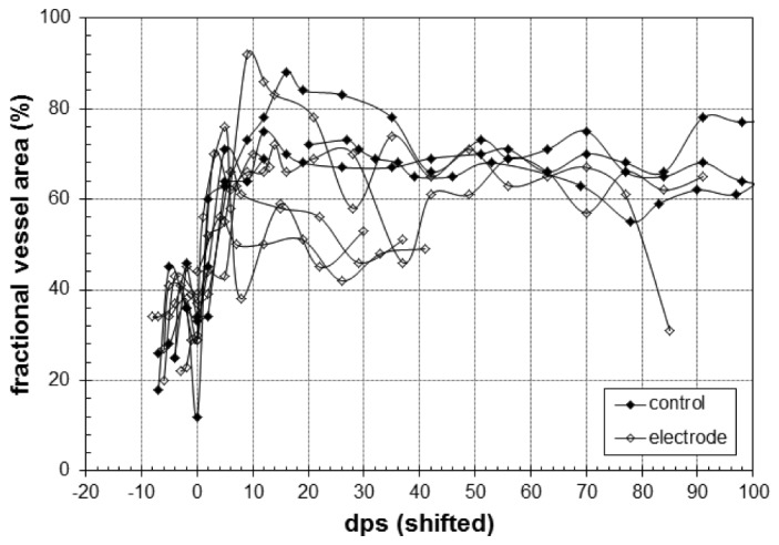
Fractional area (%) covered by vessel in first depth plane (0-100 µm) for control (closed symbols) and electrode (open symbols) animals in a 500 × 500 µm region. For visual clarity, the curves are aligned to the second minimum, which is set to day 0 (i.e., after vessel dilation and before new vessel growth).