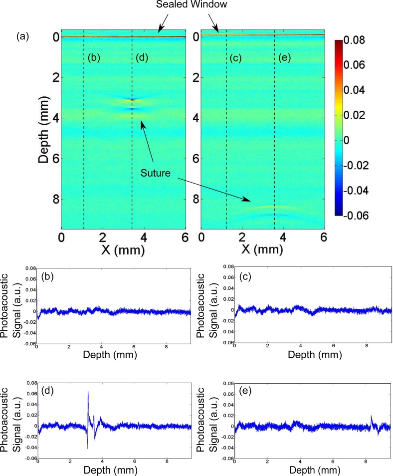
Fig. 8.
(a) Two raw 2D images showing the photoacoustic signal (a.u.) from the suture at two depths; 3.3 mm below the surface and 8.4 mm below the surface; (b)–(c) plots of the A-line of the planes without suture and (d)–(e) plots of the A-line of the planes with the signal of the suture. Quadriphasic signals that are due to the presence of a suture are distinguishable from the background noise and artifacts.

