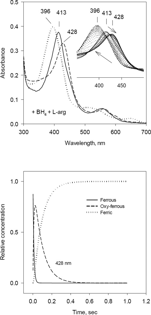
Fig. 1. Optical absorbance spectra and kinetics of the heme species observed during single turnover reaction of iNOSox(+BH4, +L-Arg) with oxygen.
Anaerobic ferrous iNOSox (8–10 µM) containing 500 µM of L-Arg and 10 µM of BH4 was rapidly mixed with an air-saturated buffer at 22 °C. The stopped-flow spectra were collected at 2.5 ms intervals using a rapid scan diode-array detector. Data are representative of three experiments.
A) UV-VIS spectra of starting ferrous Fe(II) (solid ine), oxy-ferrous intermediate Fe(II)O2 (dashed line) and final ferric Fe(III) (dotted line) species resolved by global analysis.
Inset: Original rapid-scan diode-array data. Arrows indicate directions of the spectral shift with time.
B) Kinetics of Fe(II) (solid line), Fe(II)O2 (dashed line) and Fe(III) (dotted line) resolved from global analysis. Data are expressed as percentage of the total heme concentration.

