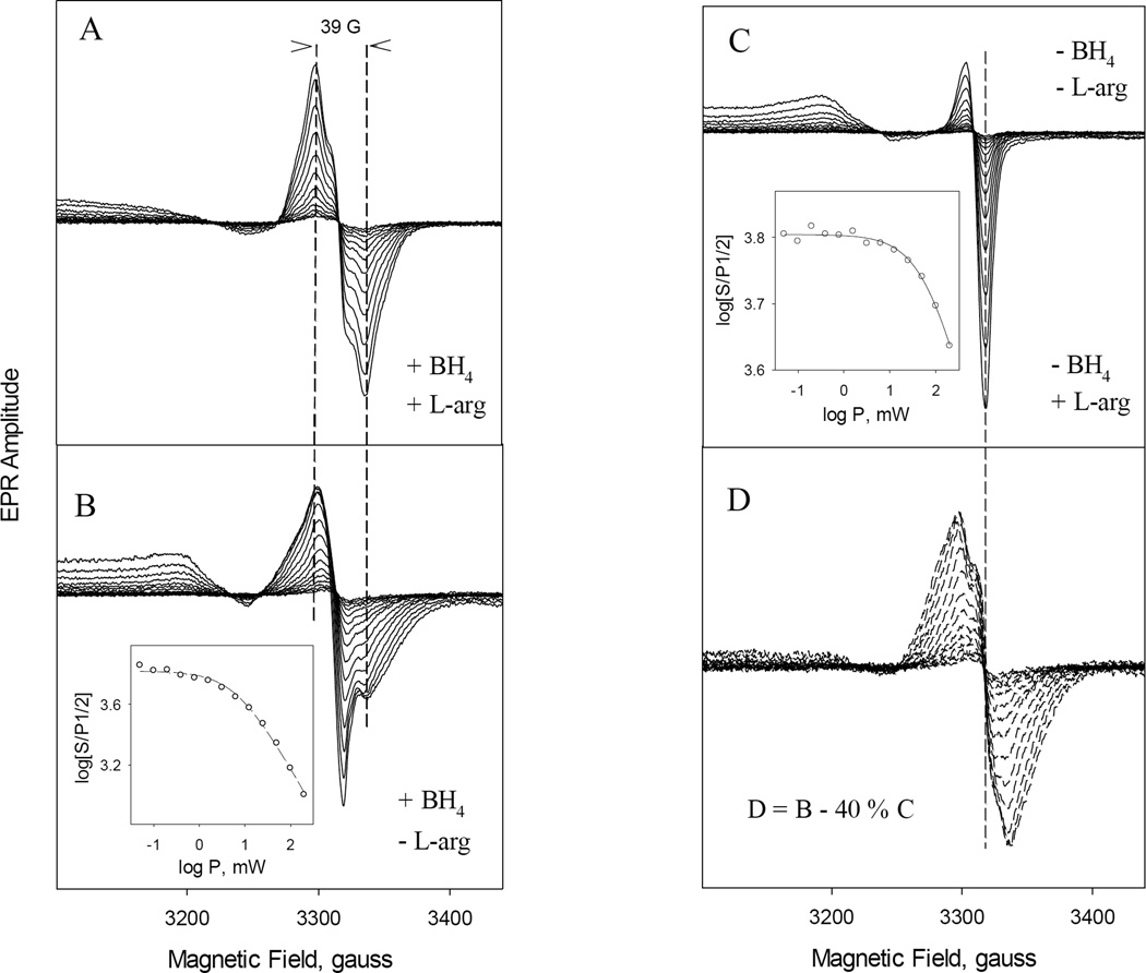
Fig. 9. Progressive power-dependent EPR spectra of the radical intermediates formed in the reaction between ferrous iNOSox and oxygen.
The reaction was identical to that described in Fig. 8. EPR spectra were recorded at microwave powers ranging from 0.025–100 mW with 3 dB increment for 40–50 µM iNOSox with equimolar concentration of BH4 and 500 µM L-Arg present (A) or with BH4 alone (B) or with L-Arg alone (C) at 115 K. Almost identical EPR spectra shown in (C) were obtained for iNOSox in the absence of both BH4 and L-Arg (data not shown). Difference spectra obtained by subtracting of 40% spectra (C) from spectra (B) to maximally remove the g= 2.005 sharp dip signal are shown at panel (D). Spectra D are very similar to typical EPR spectra of BH4 radical of iNOSox (A). Vertical dash lines are visual guides. Insets in (B) and (C) are plots to obtain the half-saturation power via non-linear regression to Equation 1. Symbols are the data and lines are the fit.

