
Figure 2. Path of the ErmBL compared to TnaC and SecM nascent chains.
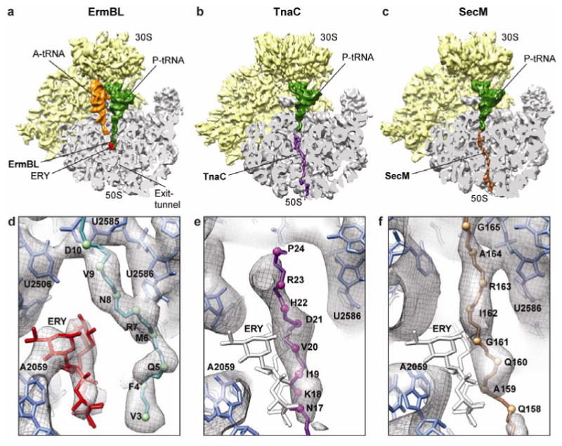
a-c, Comparison of cryo-EM structures of ErmBL-SRC, TnaC-SRC8 and SecM-SRC10, with a transverse section of the 50S subunit (grey) displaying the path of the respective nascent chains through the ribosomal tunnel. d-f, Paths of the (d) ErmBL (teal), (e) TnaC8 (purple) and (f) SecM10 (brown) nascent chains. Erythromycin (ERY)11 is colored red in (d) ErmBL-SRC, whereas the superimposed position of ERY is colored white in (e) TnaC-SRC and (f) SecM-SRC.