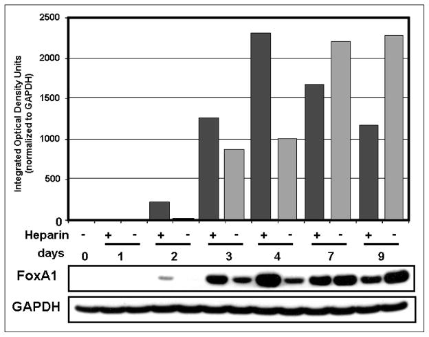
FIGURE 3.
FoxA1 protein expression is affected by heparin. FoxA1 protein in whole cell lysates of isolated human AT2 cells was analyzed by Western blot. Levels of FoxA1 steadily increased from day 2 in untreated cells, as confirmed in the densitometric tracings (gray bars). FoxA1 was highly increased by heparin treatment (black bars) at days 2, 3, and 4 but was decreased on days 7 and 9 by heparin. GAPDH of the same blot is shown as a loading control. For normalization of FoxA1 densitometry, equal areas of GAPDH signal in each lane were quantified to avoid signal overlap.

