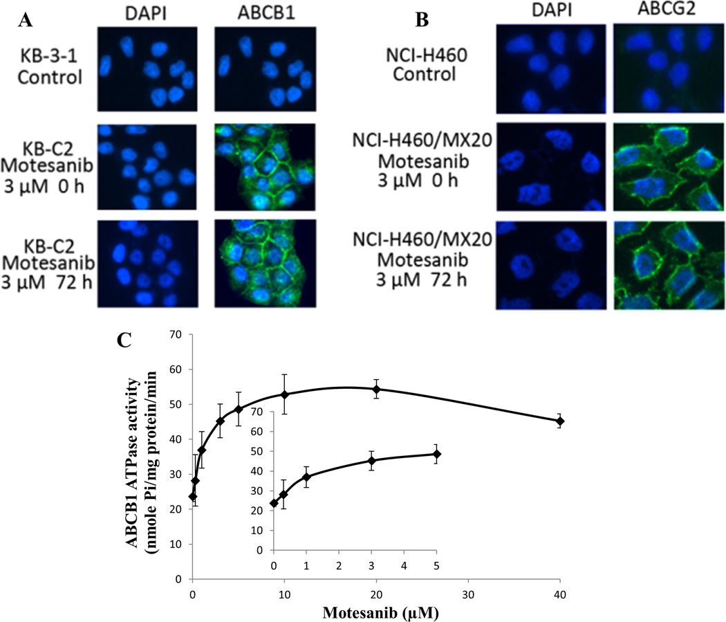
Figure 4. Effect of motesanib on the subcellular localization of ABCB1 and ABCG2, and effect of motesainib on the Vi-sensitive ABCB1 ATPase activity.
(A) Effect of motesanib at 3 µM on the subcellular localization of ABCB1 in KB-C2 cells for 72 h. (B) Effect of motesanib at 3 µM on the subcellular localization of ABCG2 in NCI-H460/MX20 cells for 72 h. ABCB1 and ABCG2 staining are shown in green. DAPI (blue) counterstains the nuclei. (C) Crude membranes (100 µg protein/ml) from High-five cells expressing ABCB1 were incubated with increasing concentrations of motesanib (0–40 µM), in the presence and absence of sodium orthovanadate (Vi) (0.3 mM), in ATPase assay buffer as described in ‘‘Materials and methods”. The inset shows stimulation of ATP hydrolysis at lower (0–5 µM) concentration of motesanib.

