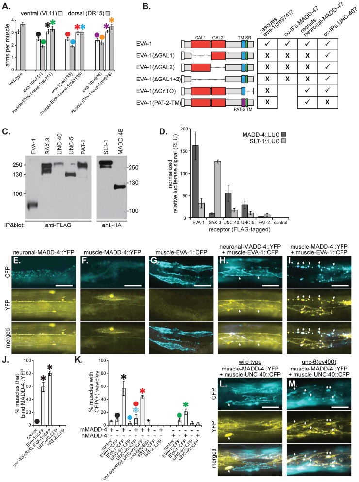
Figure 3. EVA-1 functions cell-autonomously in muscles and interacts with MADD-4.
A. Muscle-expressed EVA-1::CFP rescues the muscle extension defects of eva-1 mutants. B. A summary of EVA-1 domain function that is fully detailed in Figure S1. C&D. FLAG-tagged receptors were expressed from HEK293 cells, bathed in conditioned media from other HEK293 cells that express HA- and Gaussia luciferase-tagged MADD-4 or SLT-1 ligands, and immunoprecipitated to determine the relative amounts of ligand that co-immunoprecipitates with the receptor (see the materials and methods section for more details). C. The western blot on the left shows the five immunoprecipitated FLAG-tagged receptors. The western blot on the right shows the two HA- and Gaussia luciferase-tagged ligands that were collected from cell culture. D. The normalized relative levels of luciferase signal that immunoprecipitated with each potential ligand-receptor complex. E–I. Shown are animals harbouring one of three different transgenes that drive the expression of either neuronally-expressed MADD-4::YFP (from the trIs66 transgenic array) (E), muscle-expressed MADD-4::YFP (from the trIs78 transgenic array) (F), or muscle-expressed EVA-1::CFP (from the trIs89 transgenic array) (G), or animals harbouring two of the transgenes; trIs66 and trIs89 (H) and trIs78 and trIs89 (I). The relative levels of MADD-4::YFP expression from trIs66 and trIs78 is shown in Figure S2a. Images show either the CFP channel (top), YFP channel (middle) or a merged view (bottom). Arrows in ‘H’ indicate the localization of MADD-4::YFP to EVA-1::CFP expressing muscles; arrows in ‘I’ indicate the vesicularization of MADD-4::YFP and EVA-1::CFP in the muscle cells. J. The quantification of neuronally-secreted MADD-4::YFP localization to muscles over-expressing the indicated receptor. K. The quantification of CFP vesicles in animals that over-express the indicated CFP-tagged receptors (x-axis) in muscles in either the presence of MADD-4::YFP expressed from dorsal muscles (mMADD-4) or pan-neuronally (nMADD-4). The colocalization of MADD-4 and EVA-1 with the RAB-11 and RAB-5 endosomal markers are shown in Figure S2b and S2c. L&M. MADD-4::YFP fails to induce obvious vesicularization of UNC-40::CFP in a wild type background (L), but YFP-CFP vesicles are obvious in animals that lack UNC-6 (M). In A, J, and K, statistical significance (p<0.05) is indicated with a solid asterisk which is matched with a dot above the data point to which the comparison was made. In all micrographs, the scale bar represents 50 micrometers. In all graphs, standard error of the mean is shown.

