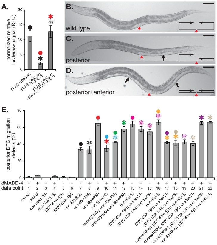
Figure 5. EVA-1 is sufficient to confer sensitivity of UNC-40 to MADD-4 in the background of UNC-6.
A. Co-expression of UNC-6 interferes with the ability of HEK293-expressed FLAG-tagged UNC-40 to co-immunoprecipitate luciferase-tagged MADD-4. Co-expression of EVA-1 relieves UNC-6's interference of the MADD-4-UNC-40 interaction. B–D. Examples of the phenotypic consequence of wild-type (B) and errant distal tip cell migration (C&D). In the micrographs, the red arrowhead indicates the vulval slit, and the black arrows indicate the white ventral patch that results from a guidance error in distal tip cell migration. The line/arrow schematics in the lower right of each micrograph chart the migratory path of the anterior and posterior distal tip cells in the respective animal. Dorsal is up and anterior is to the left. In all micrographs, the scale bar represents 50 µM. E. Distal tip cell migration defects either in the presence (+) or absence (−) of dorsal muscle-expressed (m)MADD-4 (from the trIs78 transgenic array) for the indicated genotype. For simplicity, only the posterior distal defects are shown; the anterior defects are reported in supplemental Figure S4 and show similar trends. The purpose of the ‘data point’ row is to provide clarity within main text. EVA-1::CFP was expressed in the distal tip cells from the emb-9 promoter as was previously done for UNC-5 [28]. The two extrachromosomal arrays are called trEx948#1 ([DTC-EVA-1]#1 on the graph) and trEx948#2 ([DTC-EVA-1]#2 on the graph). For each graph, statistical significance (p<0.01) is documented as described for figure 1f. Standard error of the mean is shown in both graphs.

