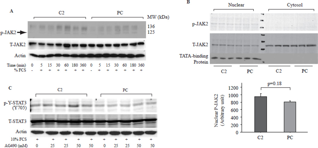
Fig. 3. JAK2 was not involved in TP induced STAT3 activation.
A. Cells treated as in Fig. 2A were subjected for Western blot assay to assess JAK2 activation and expression. P-JAK2 indicates phosphorylated Jak2, T-Jak2 indicates total JAK, actin was used as loading control. B. Western blot assay of JAK2 in the nuclear and cytosolic fractions of C2 and PC cells as prepared in Fig. 2C. TATA-binding protein was used as purity control. Bar graphs showed cumulative data of Western blot band densities of nuclear p-JAK2. N=3. C. Synchronized cells were stimulated with 10% FCS in the presence of AG490, a JAK2 inhibitor for 1 hr and then the cell lysates were subjected for Western blot assay with antibodies to p-Y-STAT3 and T-STAT3, Blot for actin was used as loading control.

