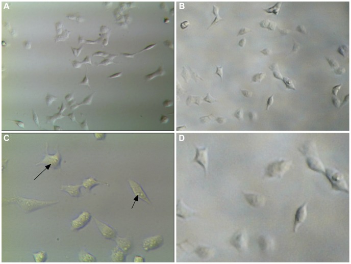
Figure 6. Comparison of MDA-MB-231 cell images between our system and a conventional optical microscope.
(a) 310× image taken at 640×480 with a conventional inverted microscope; (b) 206.8× image taken at 640×480 with our system and enlarged post acquisition by 149% to match the size; (c) 620× image taken at 1280×960 with a conventional inverted microscope, arrows show intra-cellular detail; (d) 1280×960 image taken with our system at full magnification (413.6×) and enlarged post-aquisition by 149% to match the size.

