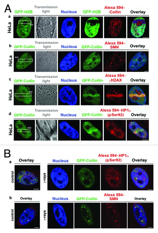
Figure 7. Visualization of selected proteins localized to DNA lesions. (A) (a) Recruitment of endogenous coilin (red) to DNA lesions was studied in HeLa cells stably expressing histone H2B (green). (b) GFP-coilin (green), but not other CB-related protein, called SMN, was recruited to UVA-damaged chromatin. (c) High levels of GFP-coilin (green) and γ-H2AX (red) co-localized with DNA lesions in BrdU pre-sensitized cells. (d) GFP-coilin (green) and phosphorylated HP1α at serine 92 (red) were recruited to DNA lesions in BrdU pre-sensitized cells. (B) Nuclear localization patterns of (a) phosphorylated HP1α at serine 92 and (b) SMN were identical in non-irradiated cells and cells irradiated with 5 Gy of γ-rays.
