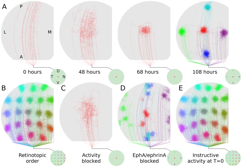
Figure 2. Retinotopic refinement.
A, Time-lapse development of a group of axons from a specific retinal location. The retina is represented by the circle at lower-right and the colored dot(s) indicate the location of RGCs whose axons are displayed in the colliculus. Development was regulated only by molecular guidance for the first 48 hours whereafter activity-dependent feedback (i.e., regulated trophic release) contributed to simulated development over the next 60 hours. Axon arbors from five retinal locations are shown in the last frame. B, Arborizations from 21 points on the retina show retinotopic order in axonal projections. Developmental paradigm is identical to ( ).
).  , Axon development over 144 hours driven only by molecular guidance. Activity-dependent feedback was required for refinement.
  , Developmental sequence as in (
 ), but with molecular guidance blocked along nasal-temporal retinal axis; anterior-posterior collicular axis. Organization is significantly disrupted.
  , Axon arbor development when activity-dependent feedback contributed to development from the time when axons first began interstitial branching (i.e., T = 0 hr in A). Development was qualitatively normal and not dominated by ectopic projections as previously predicted [36], indicating that molecular guidance and activity-dependent mechanisms are able to simultaneously guide and refine development. Data from a single simulation of retinocollicular development are shown in each of (A)-(E). In all results presented here and elsewhere, three or more simulations were performed (typical runtime 3 days each) and no qualitative differences were observed.

